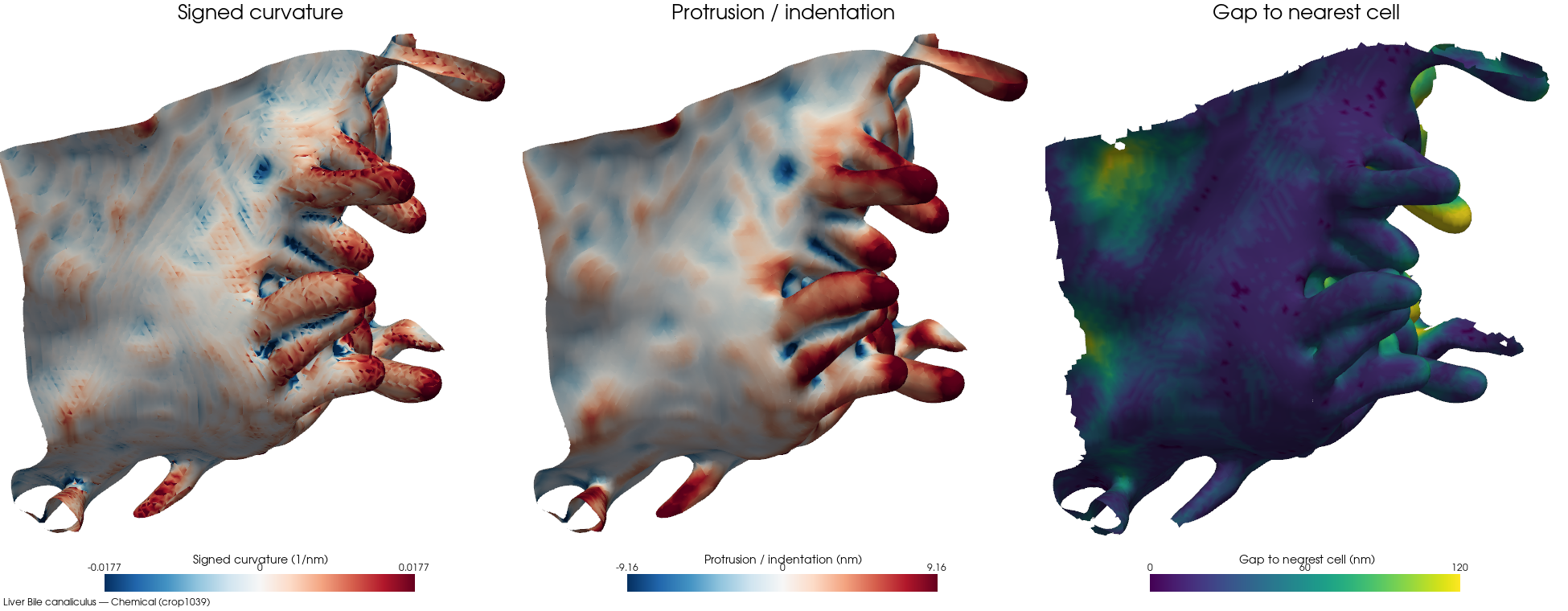
Chemicalcrop1039bd-clip 2%cell 1 · ECS frac 0.1914 · patch 62,970/115,432 faces (54% ECS-facing) · 19 cells · 16 nm · Bile canaliculus
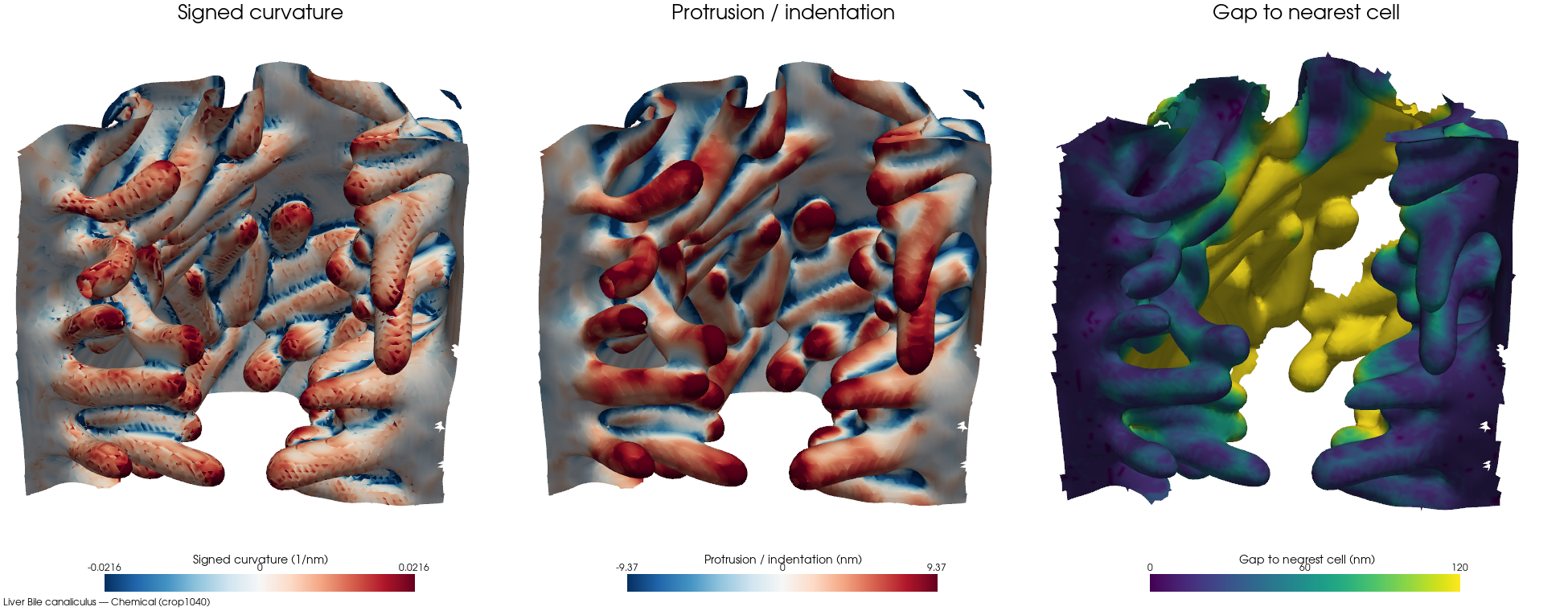
Chemicalcrop1040bd-clip 11%cell 1 · ECS frac 0.223 · patch 96,097/177,256 faces (54% ECS-facing) · 27 cells · 16 nm · Bile canaliculus
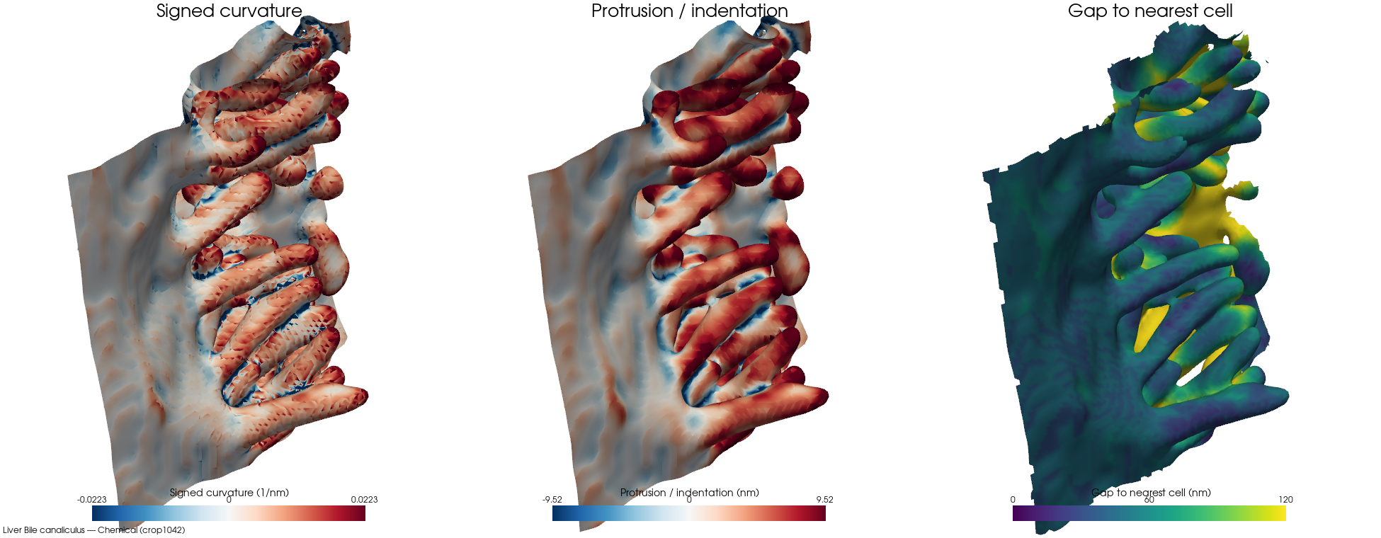
Chemicalcrop1042bd-clip 10%cell 1 · ECS frac 0.1742 · patch 80,631/154,724 faces (52% ECS-facing) · 24 cells · 16 nm · Bile canaliculus
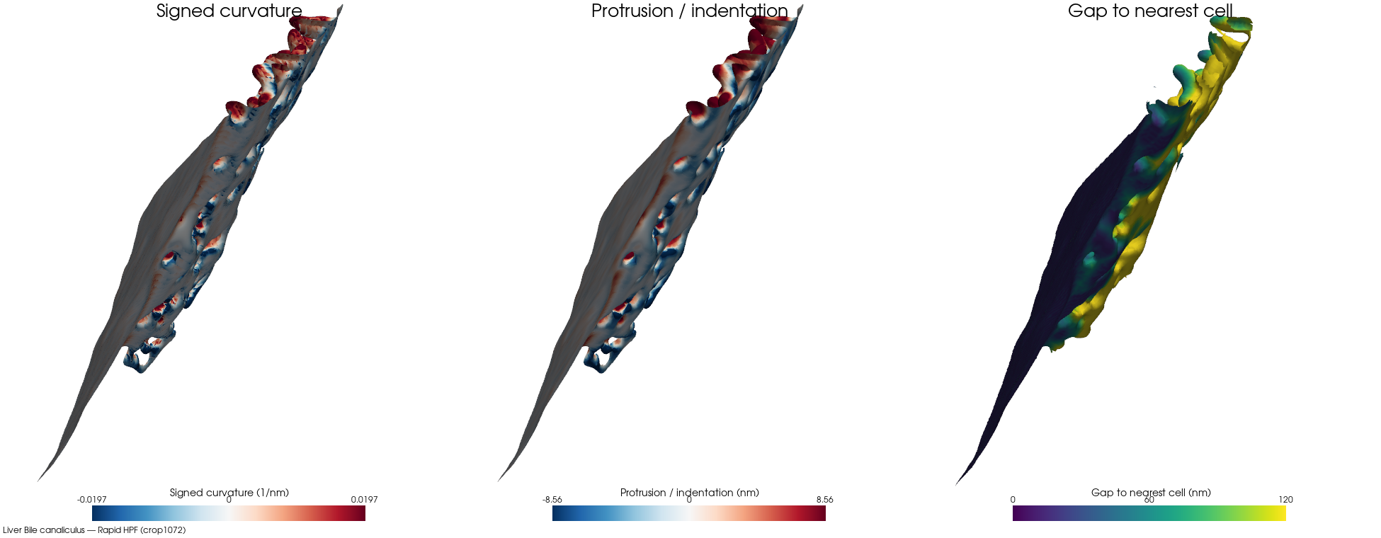
Rapid HPFcrop1072bd-clip 15%cell 1 · ECS frac 0.039 · patch 83,069/294,538 faces (28% ECS-facing) · 20 cells · 16 nm · Bile canaliculus
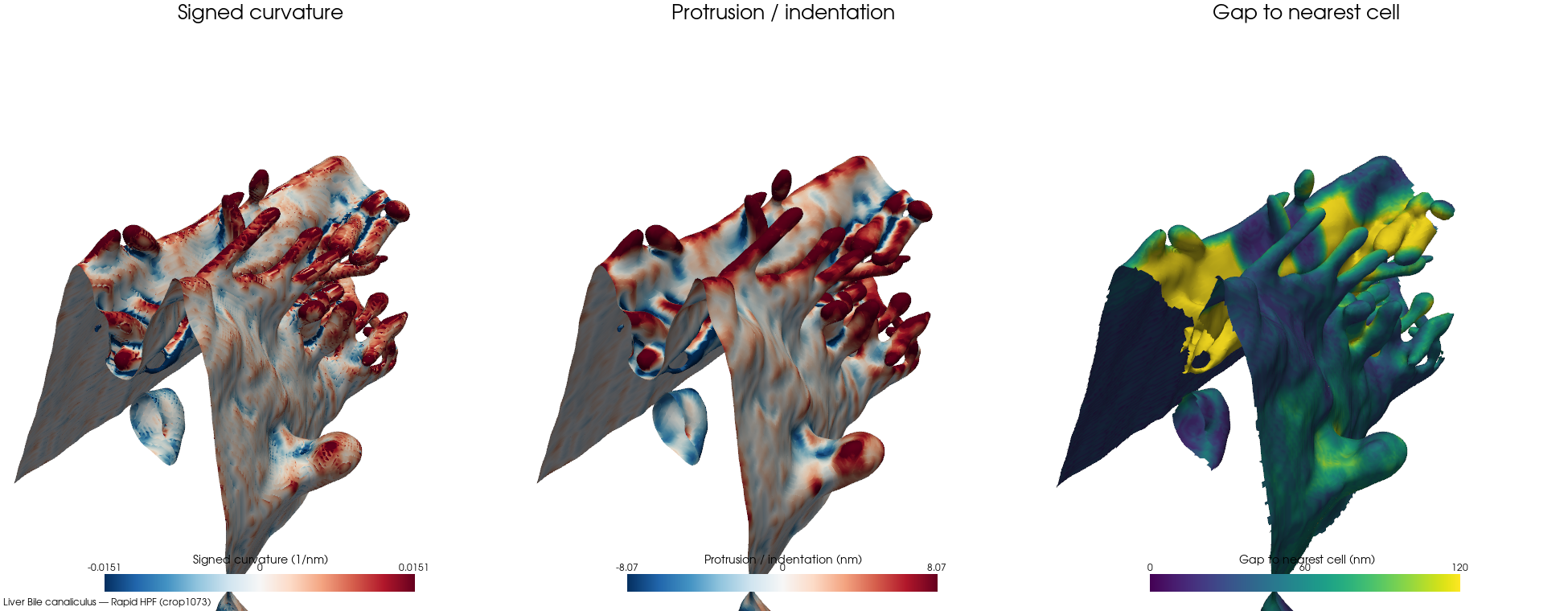
Rapid HPFcrop1073bd-clip 5%cell 2 · ECS frac 0.1416 · patch 165,510/220,248 faces (75% ECS-facing) · 32 cells · 16 nm · Bile canaliculus
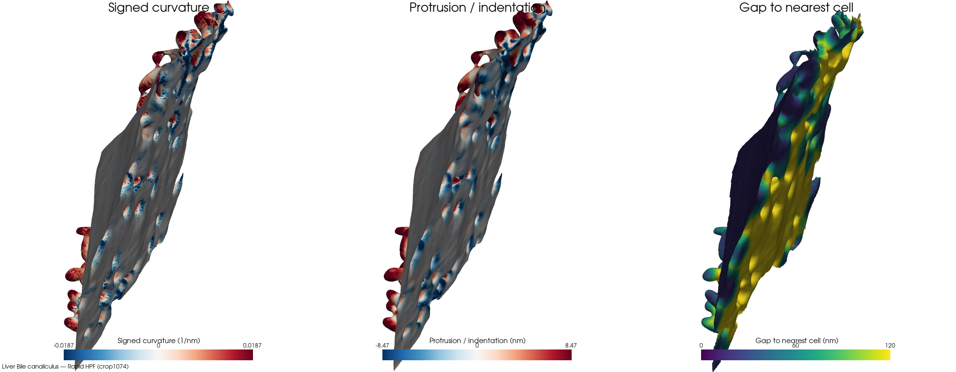
Rapid HPFcrop1074bd-clip 7%cell 1 · ECS frac 0.1014 · patch 103,430/295,332 faces (35% ECS-facing) · 44 cells · 16 nm · Bile canaliculus
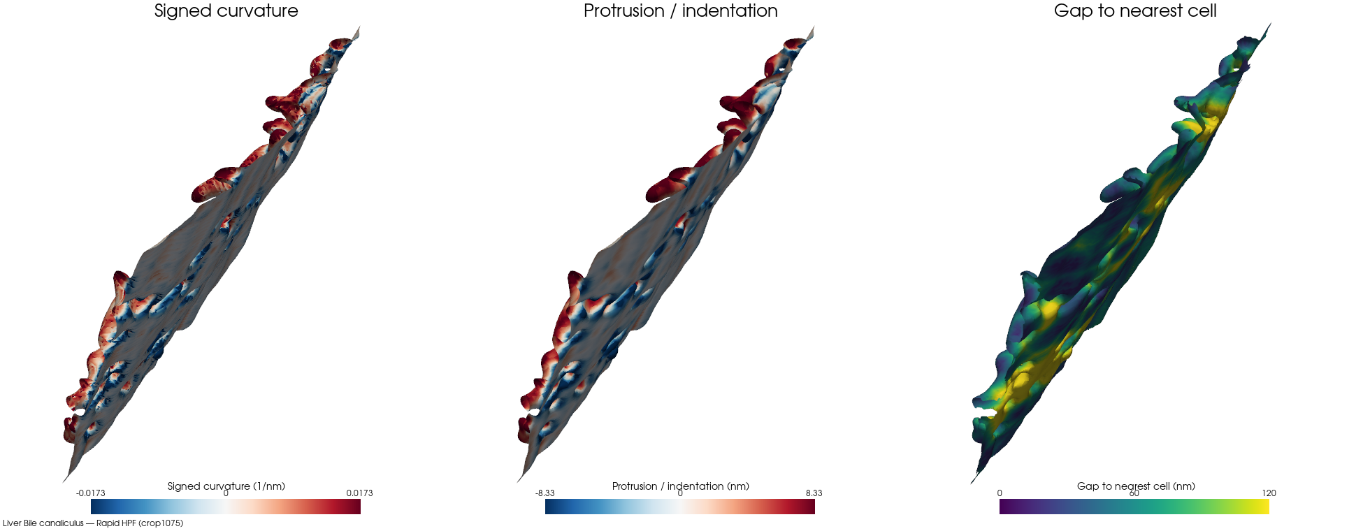
Rapid HPFcrop1075bd-clip 4%cell 1 · ECS frac 0.0448 · patch 99,083/280,264 faces (35% ECS-facing) · 55 cells · 16 nm · Bile canaliculus
Hepatocyte lateral
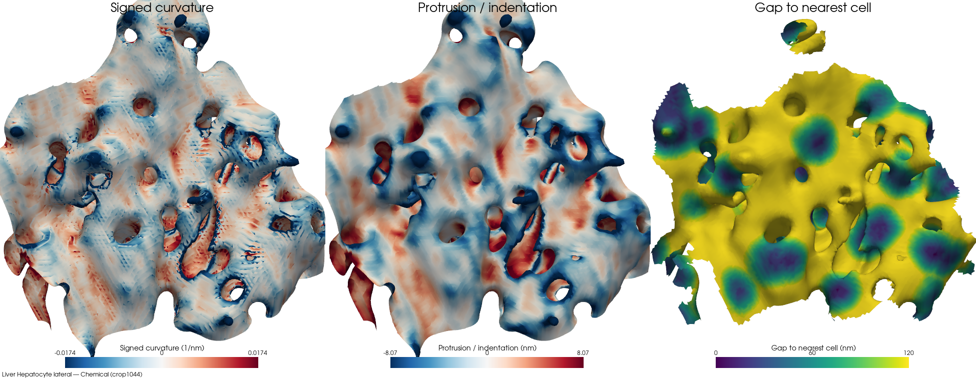
Chemicalcrop1044⚠ outlierbd-clip 23%cell 2 · ECS frac 0.2179 · patch 102,152/188,432 faces (54% ECS-facing) · 41 cells · 16 nm · Hepatocyte lateral
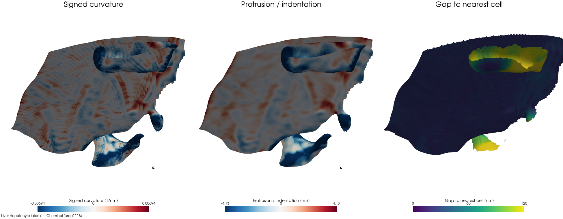
Chemicalcrop1118⚠ outlierbd-clip 15%cell 1 · ECS frac 0.0467 · patch 34,401/91,328 faces (37% ECS-facing) · 3 cells · 16 nm · Hepatocyte lateral
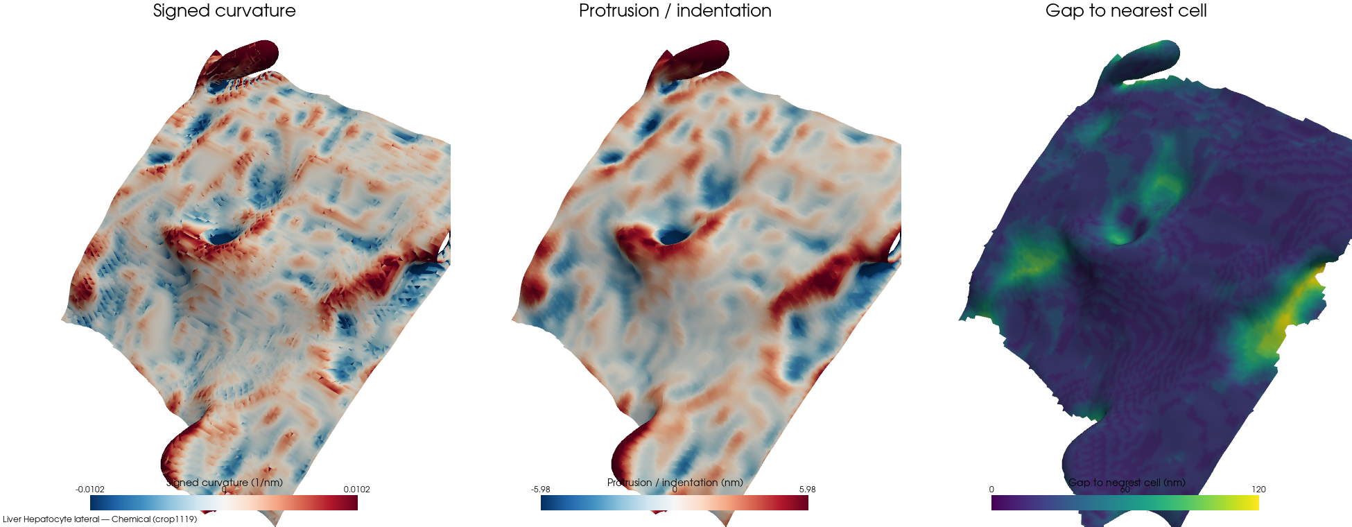
Chemicalcrop1119bd-clip 22%cell 1 · ECS frac 0.0436 · patch 36,853/77,816 faces (47% ECS-facing) · 8 cells · 16 nm · Hepatocyte lateral
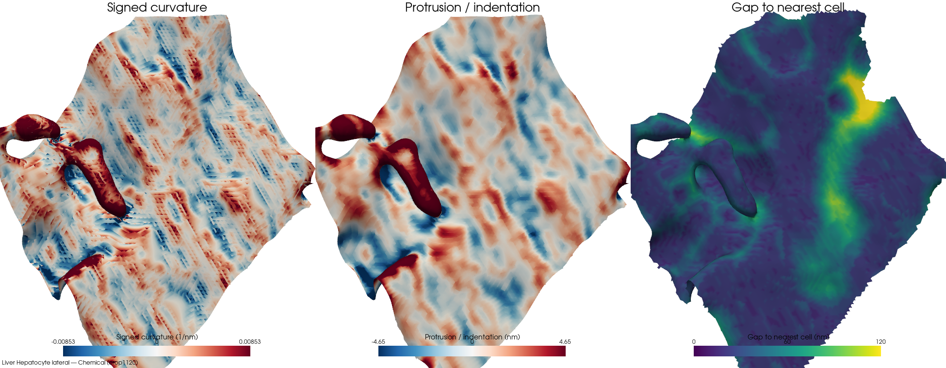
Chemicalcrop1120bd-clip 4%cell 1 · ECS frac 0.0471 · patch 36,766/93,592 faces (39% ECS-facing) · 16 cells · 16 nm · Hepatocyte lateral
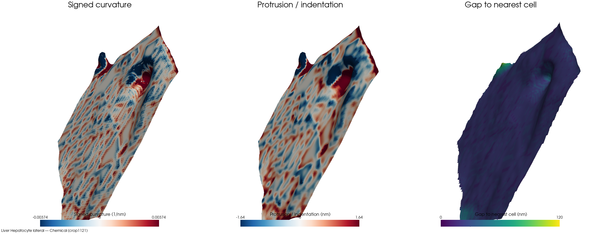
Chemicalcrop1121⚠ outlierbd-clip 1%cell 3 · ECS frac 0.0151 · patch 39,789/108,580 faces (36% ECS-facing) · 3 cells · 16 nm · Hepatocyte lateral
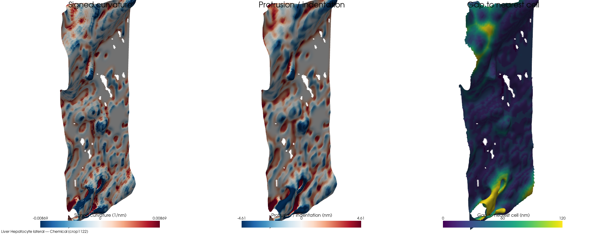
Chemicalcrop1122bd-clip 5%cell 2 · ECS frac 0.0307 · patch 40,354/127,044 faces (31% ECS-facing) · 26 cells · 16 nm · Hepatocyte lateral
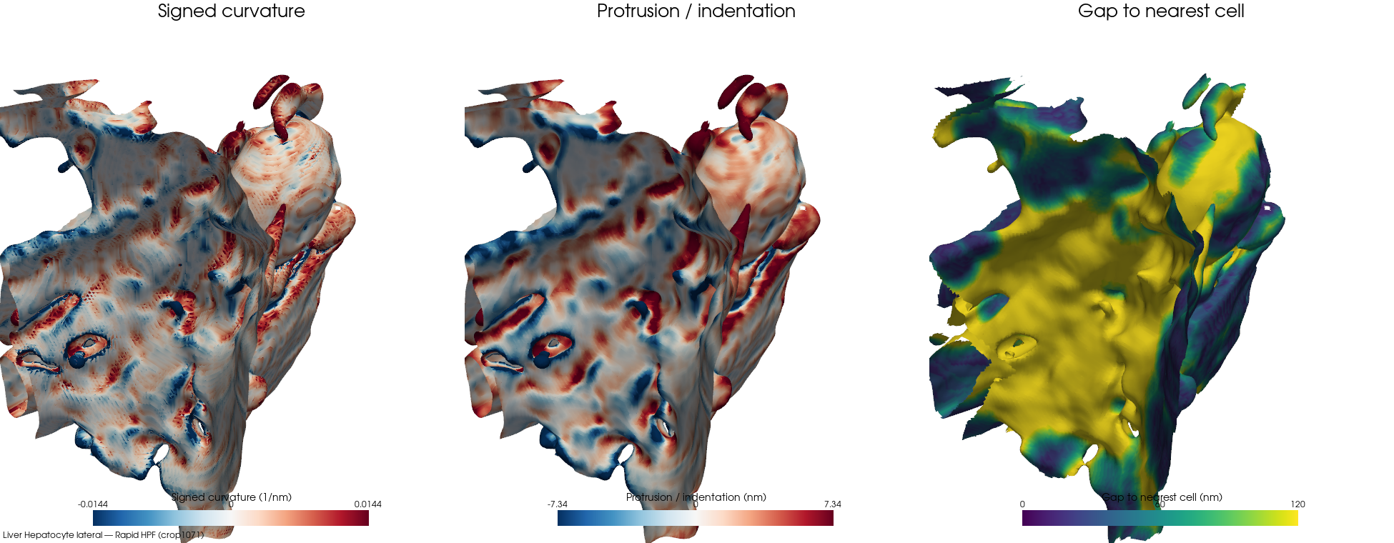
Rapid HPFcrop1071⚠ outlierbd-clip 11%cell 2 · ECS frac 0.2796 · patch 360,072/472,930 faces (76% ECS-facing) · 108 cells · 16 nm · Hepatocyte lateral
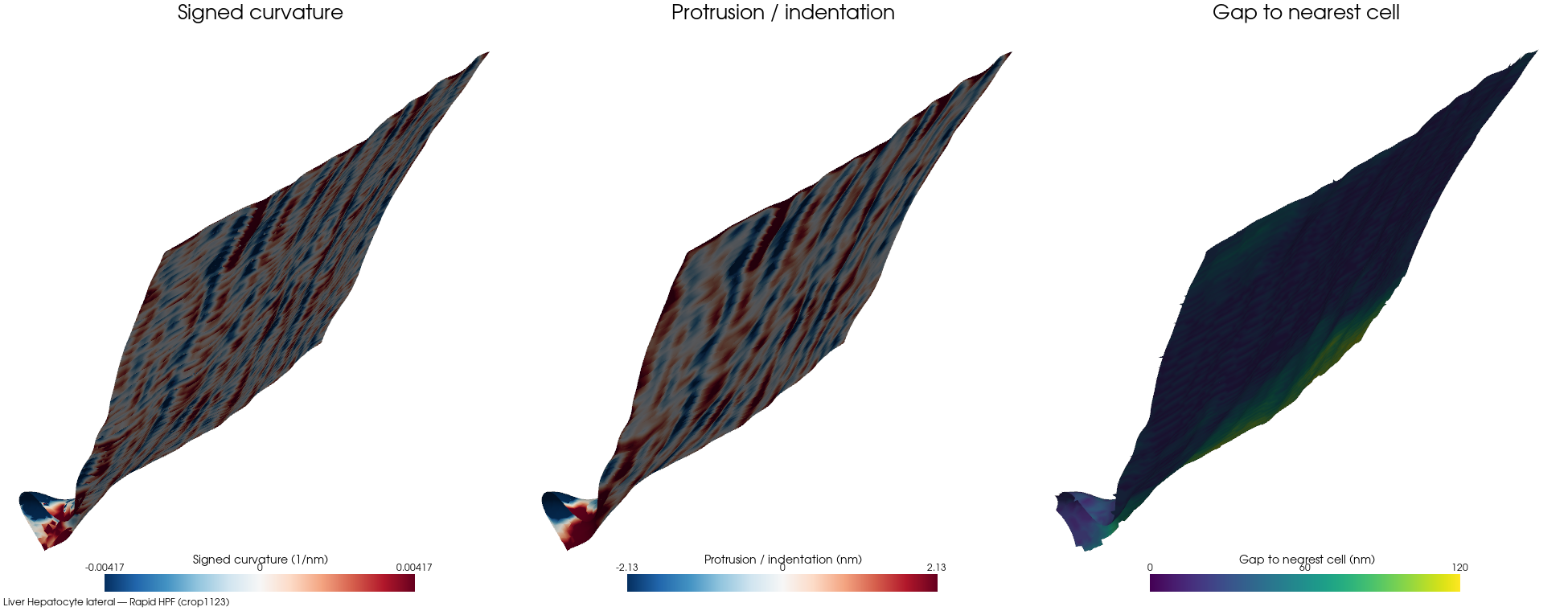
Rapid HPFcrop1123bd-clip 5%cell 1 · ECS frac 0.0253 · patch 28,893/88,940 faces (32% ECS-facing) · 3 cells · 16 nm · Tight junction
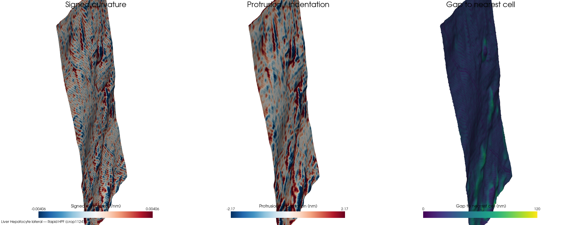
Rapid HPFcrop1124bd-clip 0%cell 2 · ECS frac 0.0193 · patch 105,775/261,500 faces (40% ECS-facing) · 10 cells · 16 nm · Hepatocyte lateral
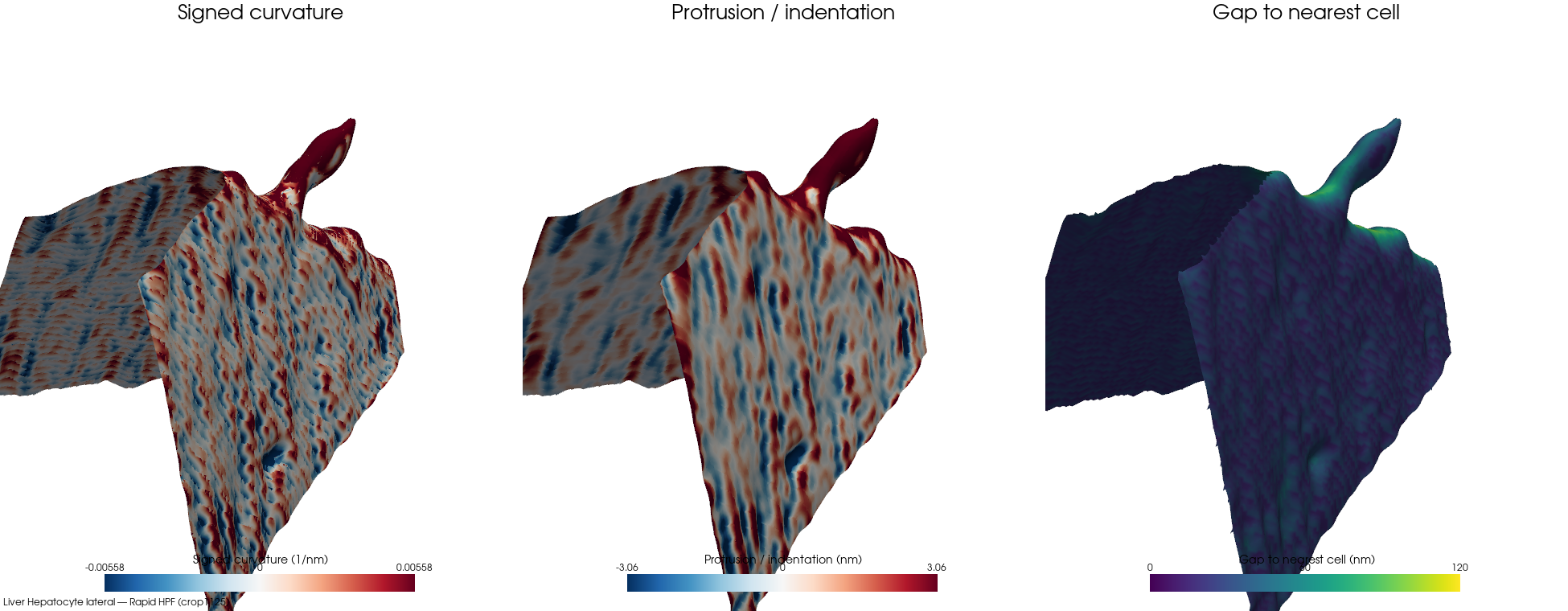
Rapid HPFcrop1125bd-clip 0%cell 2 · ECS frac 0.0313 · patch 60,008/97,908 faces (61% ECS-facing) · 3 cells · 16 nm · Hepatocyte lateral
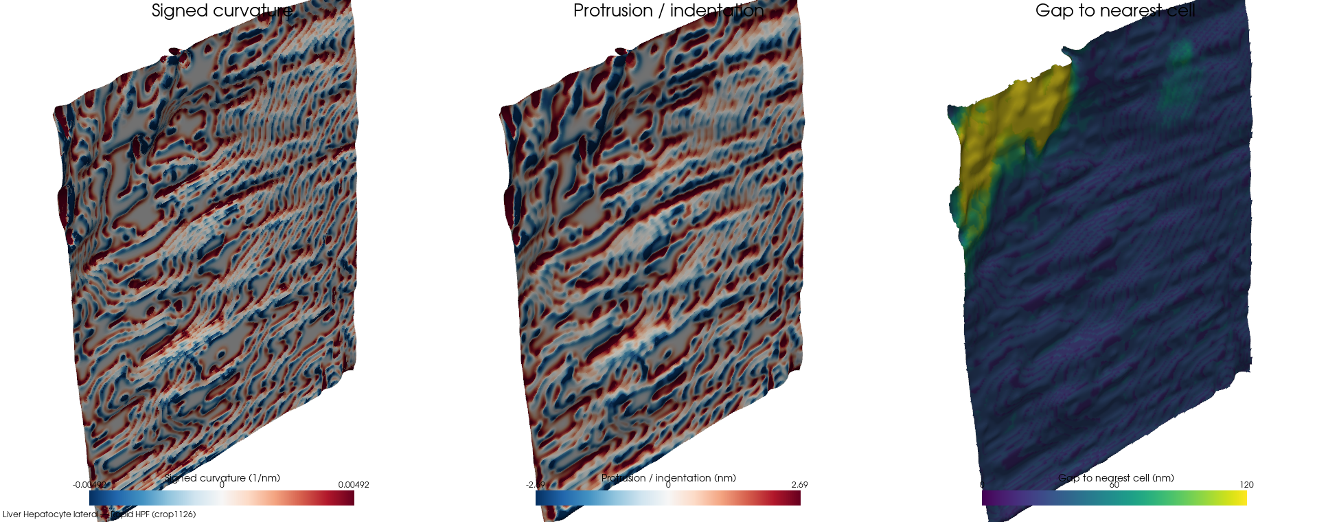
Rapid HPFcrop1126bd-clip 2%cell 2 · ECS frac 0.0211 · patch 121,784/279,540 faces (43% ECS-facing) · 2 cells · 16 nm · Tight junction
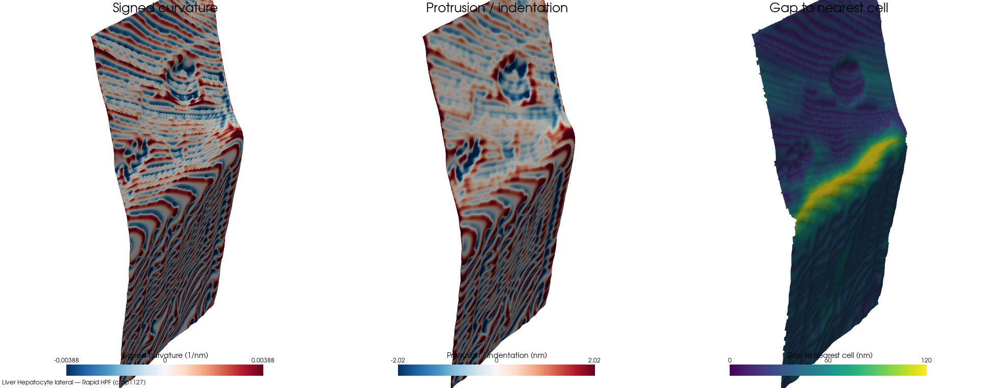
Rapid HPFcrop1127bd-clip 1%cell 1 · ECS frac 0.0398 · patch 54,519/155,920 faces (35% ECS-facing) · 8 cells · 16 nm · Hepatocyte lateral
Space of Disse
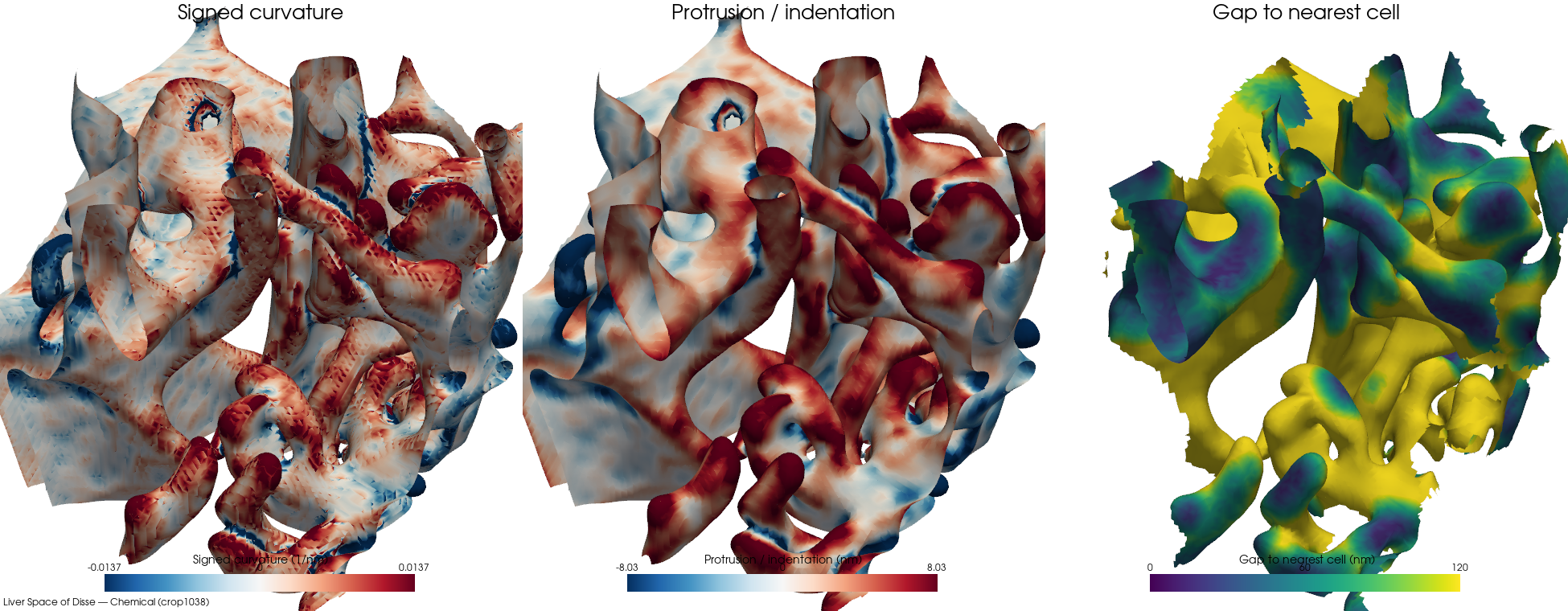
Chemicalcrop1038bd-clip 35%cell 2 · ECS frac 0.5485 · patch 180,024/234,402 faces (76% ECS-facing) · 61 cells · 16 nm · Space of Disse
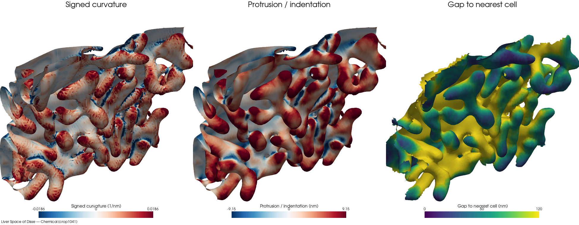
Chemicalcrop1041bd-clip 18%cell 1 · ECS frac 0.2688 · patch 108,057/178,240 faces (60% ECS-facing) · 50 cells · 16 nm · Space of Disse
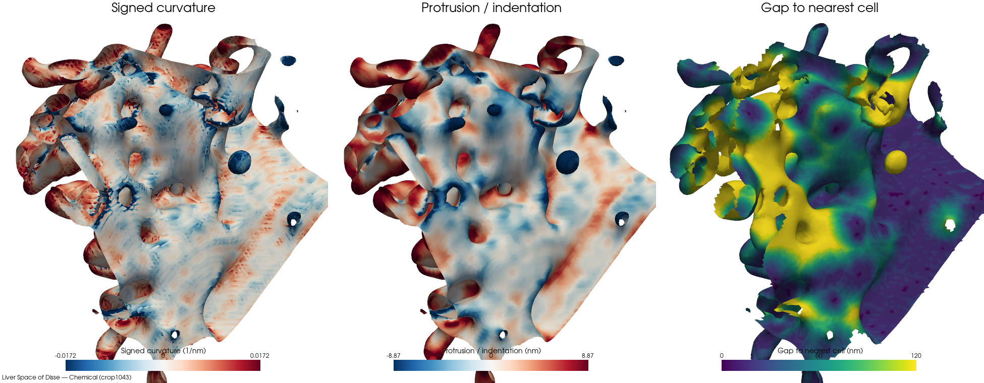
Chemicalcrop1043bd-clip 11%cell 5 · ECS frac 0.2642 · patch 110,817/152,436 faces (72% ECS-facing) · 81 cells · 16 nm · Space of Disse
Heart
Cardiac interstitial
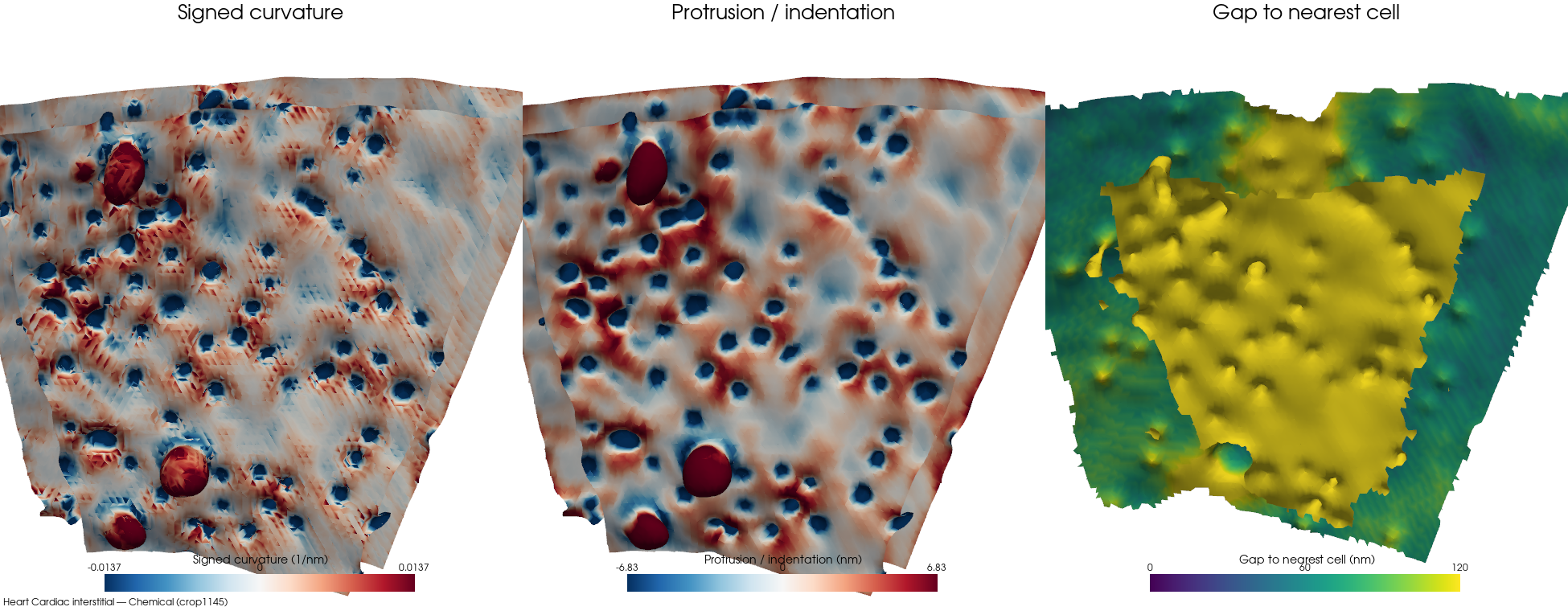
Chemicalcrop1145bd-clip 33%cell 2 · ECS frac 0.3149 · patch 64,771/79,376 faces (81% ECS-facing) · 3 cells · 16 nm · Interstitial
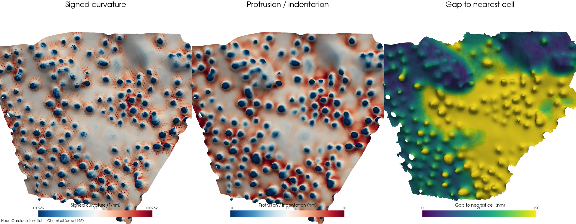
Chemicalcrop1146bd-clip 19%cell 4 · ECS frac 0.2012 · patch 44,936/96,314 faces (46% ECS-facing) · 5 cells · 16 nm · Interstitial
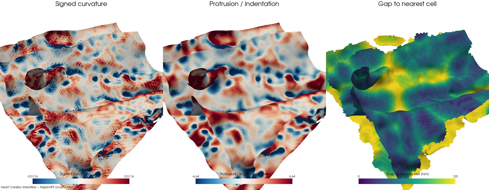
Rapid HPFcrop1149bd-clip 18%cell 2 · ECS frac 0.2126 · patch 60,829/80,096 faces (75% ECS-facing) · 31 cells · 16 nm · Interstitial
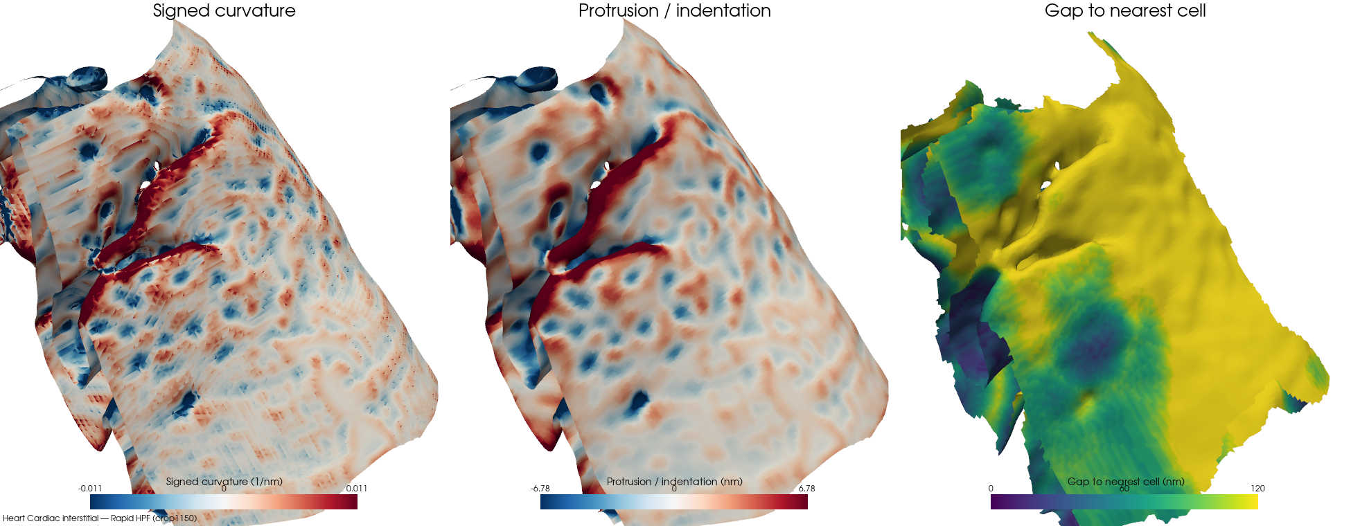
Rapid HPFcrop1150bd-clip 19%cell 3 · ECS frac 0.3839 · patch 101,052/126,500 faces (79% ECS-facing) · 33 cells · 16 nm · Interstitial
Intercalated disc
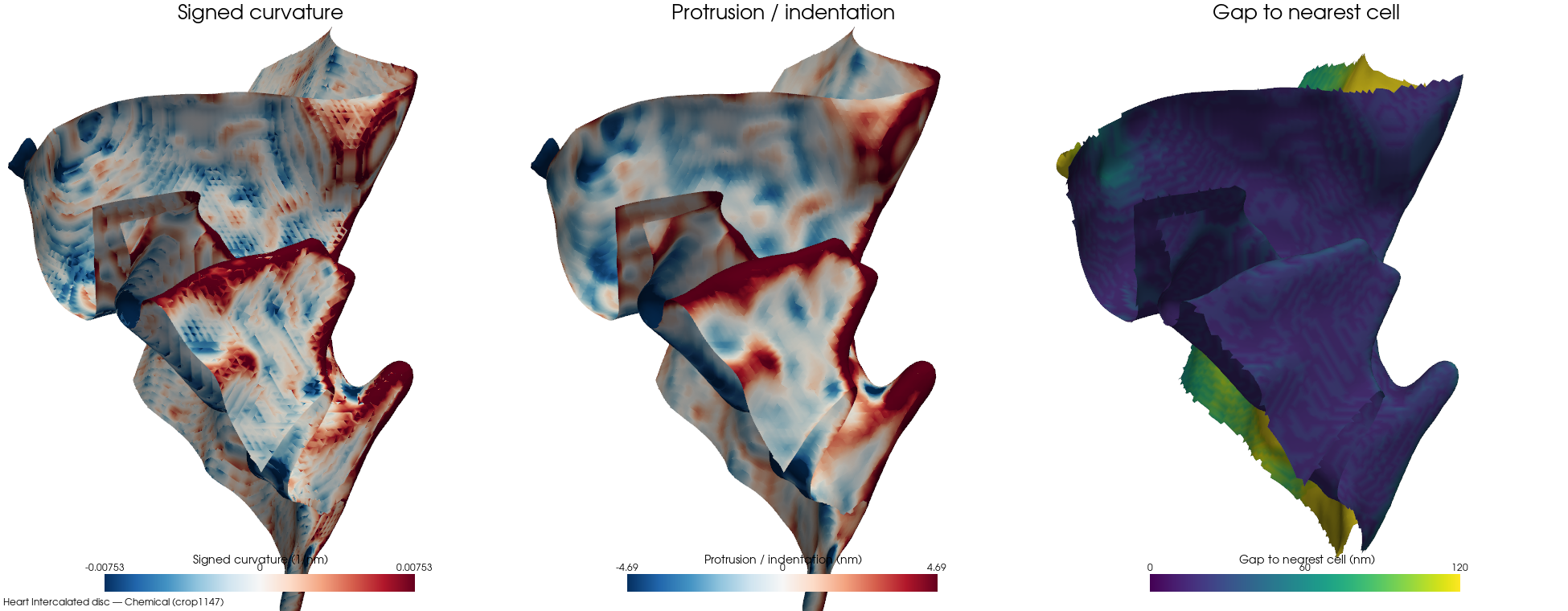
Chemicalcrop1147bd-clip 8%cell 2 · ECS frac 0.1121 · patch 62,354/114,504 faces (54% ECS-facing) · 3 cells · 16 nm · Intercalated disc
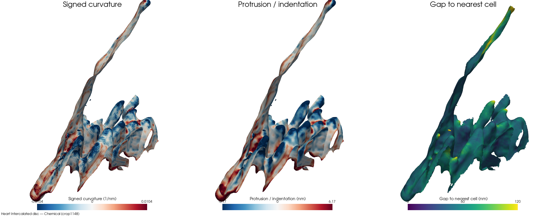
Chemicalcrop1148bd-clip 6%cell 2 · ECS frac 0.0908 · patch 141,304/237,712 faces (59% ECS-facing) · 5 cells · 16 nm · Intercalated disc
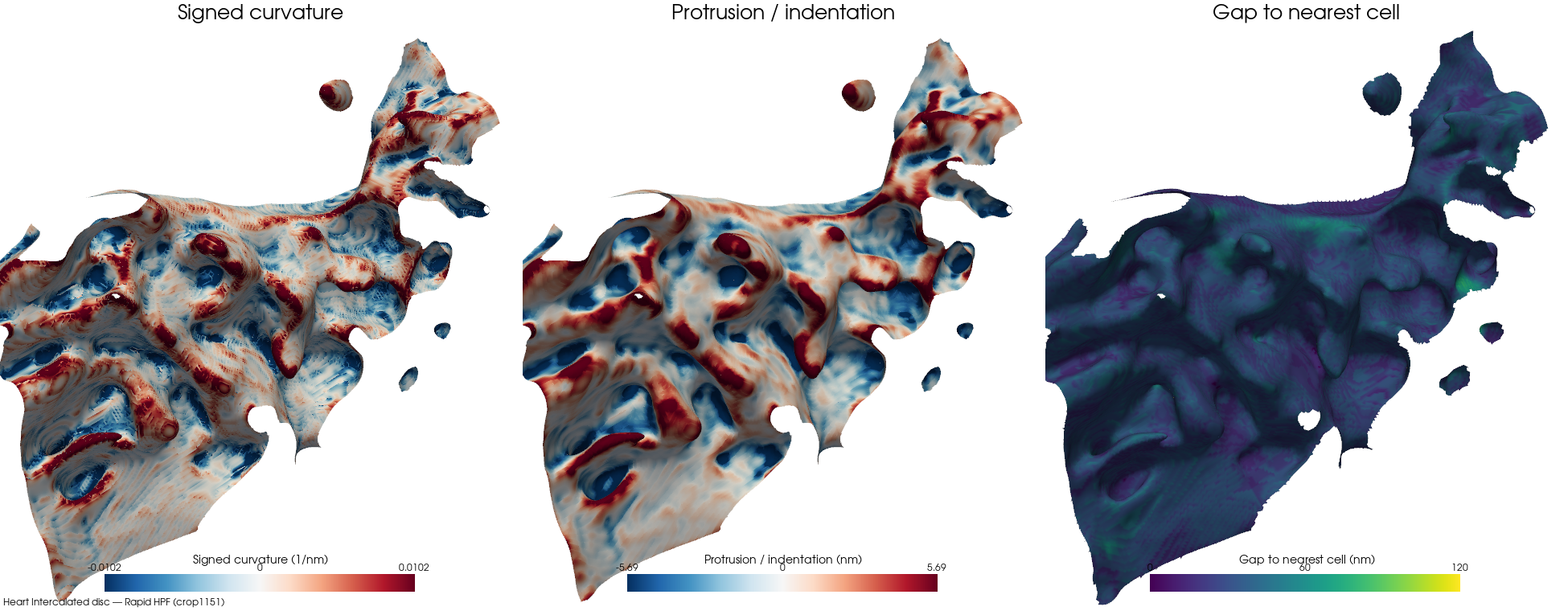
Rapid HPFcrop1151bd-clip 1%cell 1 · ECS frac 0.0452 · patch 138,389/271,848 faces (50% ECS-facing) · 5 cells · 16 nm · Intercalated disc
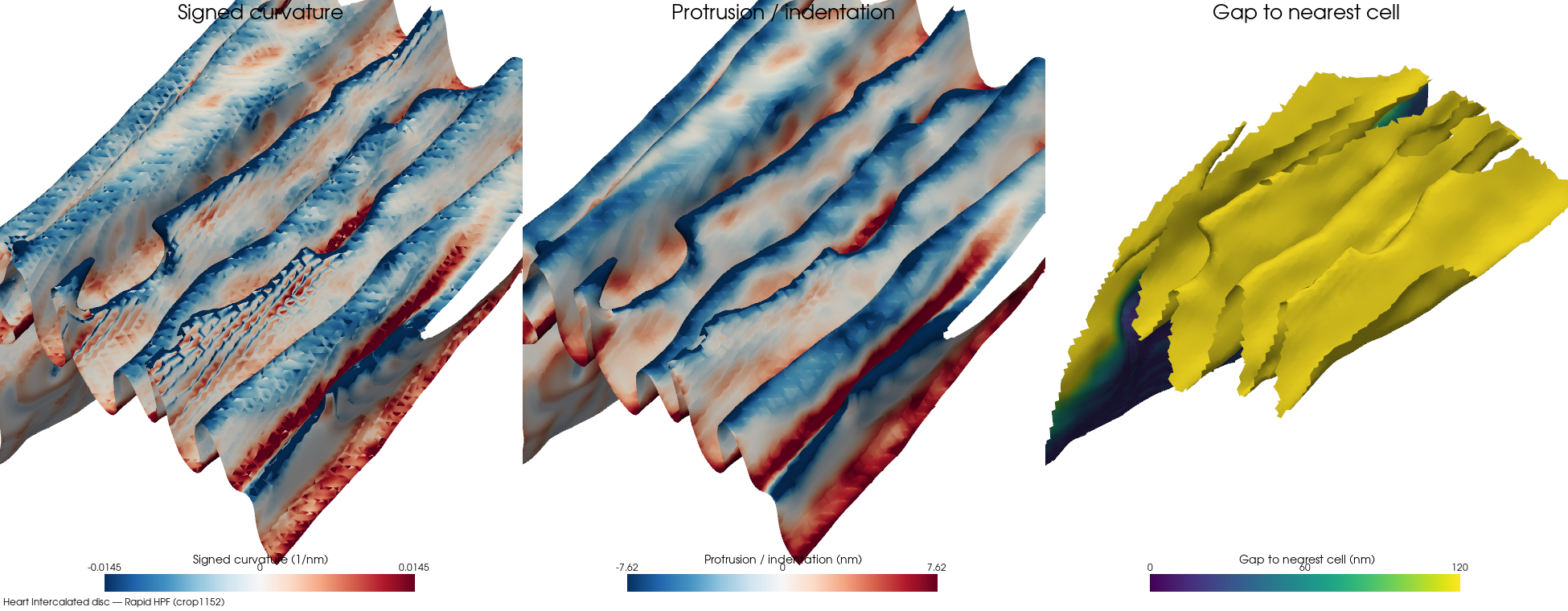
Rapid HPFcrop1152bd-clip 52%cell 1 · ECS frac 0.2 · patch 82,682/154,338 faces (53% ECS-facing) · 3 cells · 16 nm · Intercalated disc
Kidney
DCT base
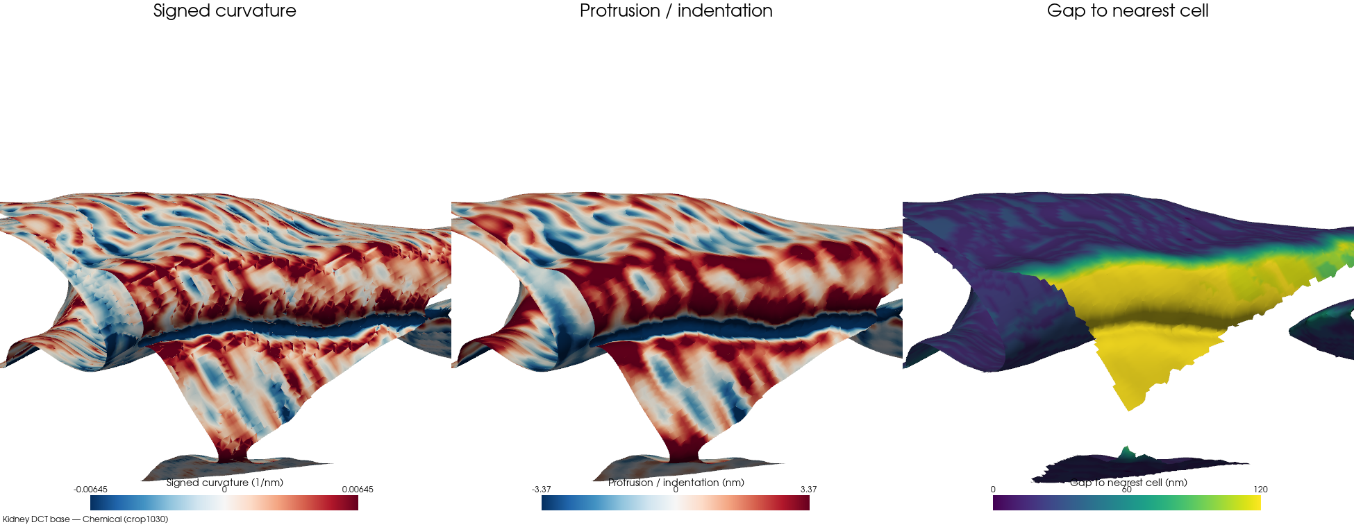
Chemicalcrop1030bd-clip 7%cell 9 · ECS frac 0.1945 · patch 61,003/96,876 faces (63% ECS-facing) · 28 cells · 16 nm · DCT base
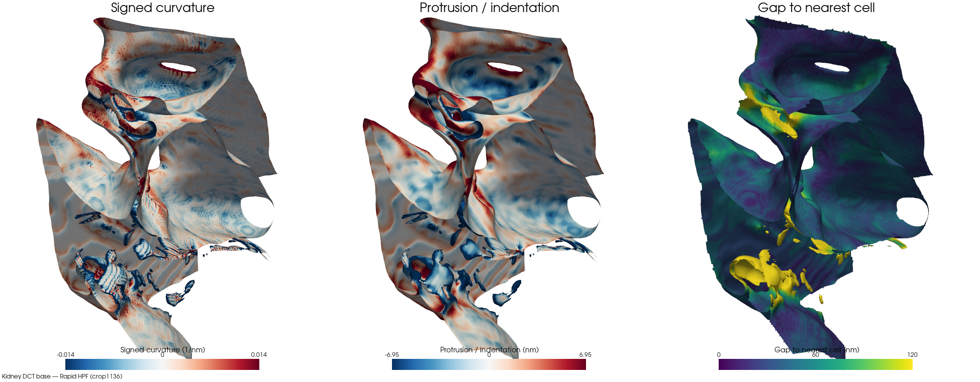
Rapid HPFcrop1136bd-clip 7%cell 10 · ECS frac 0.0894 · patch 192,564/255,502 faces (75% ECS-facing) · 19 cells · 16 nm · DCT base
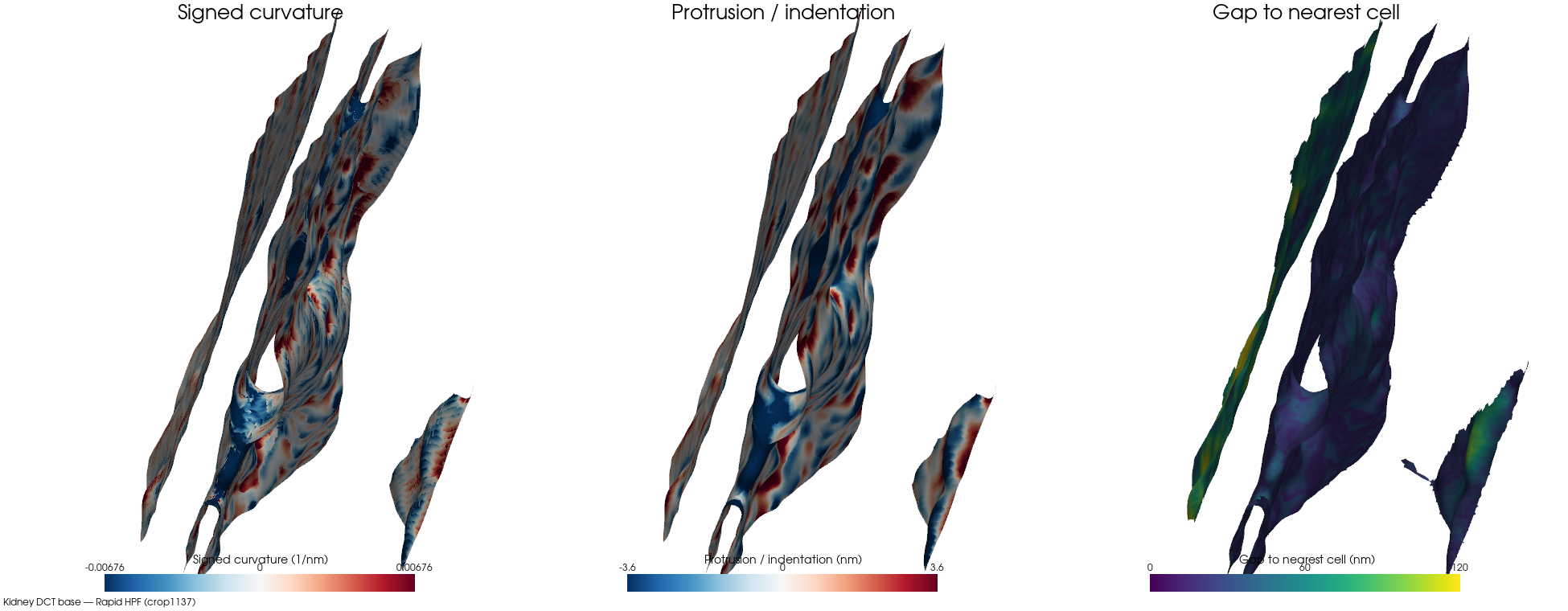
Rapid HPFcrop1137bd-clip 3%cell 1 · ECS frac 0.1469 · patch 105,613/177,208 faces (59% ECS-facing) · 31 cells · 16 nm · DCT base
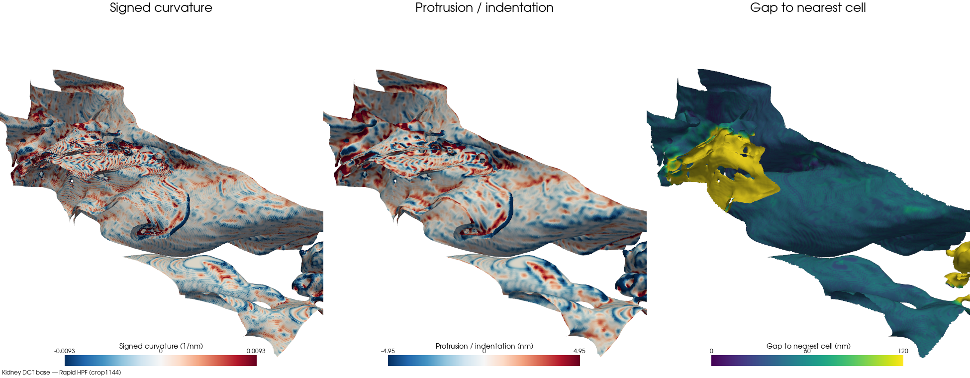
Rapid HPFcrop1144bd-clip 27%cell 1 · ECS frac 0.109 · patch 622,320/887,330 faces (70% ECS-facing) · 31 cells · 16 nm · DCT base
Glomerular
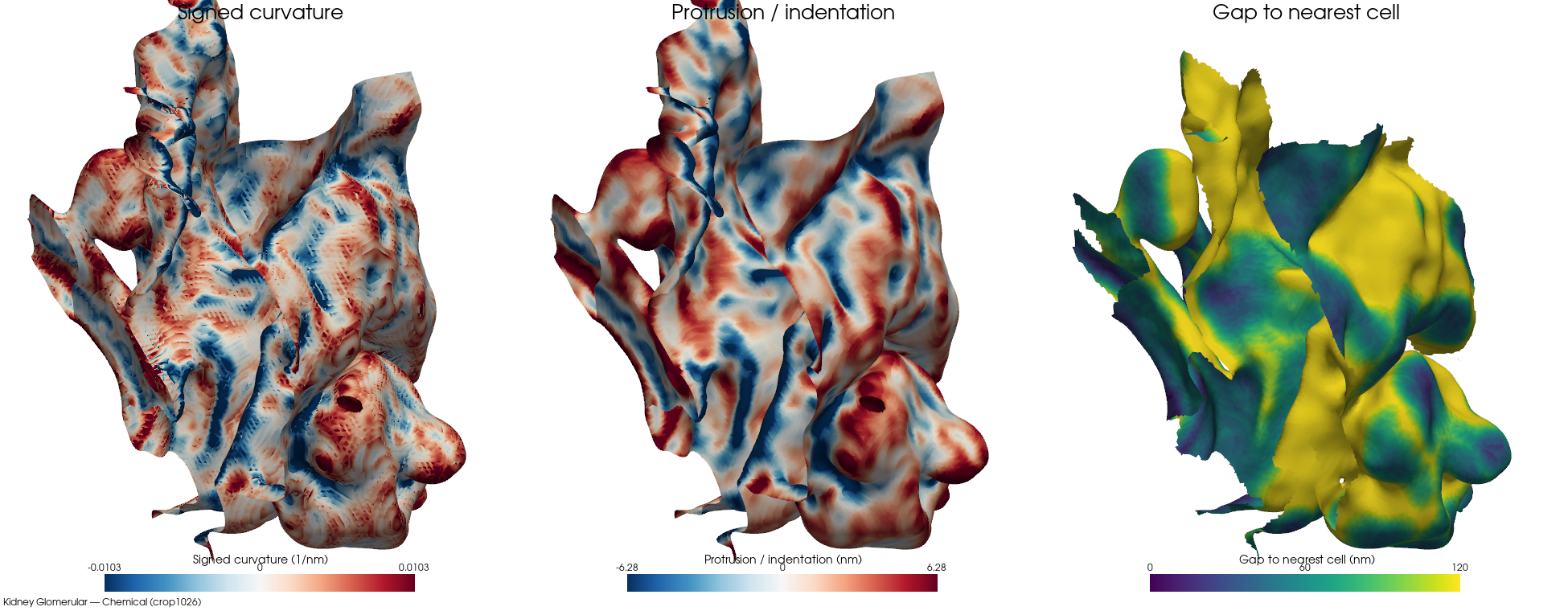
Chemicalcrop1026bd-clip 18%cell 6 · ECS frac 0.355 · patch 162,776/220,160 faces (73% ECS-facing) · 48 cells · 16 nm · Glomerular interstitium
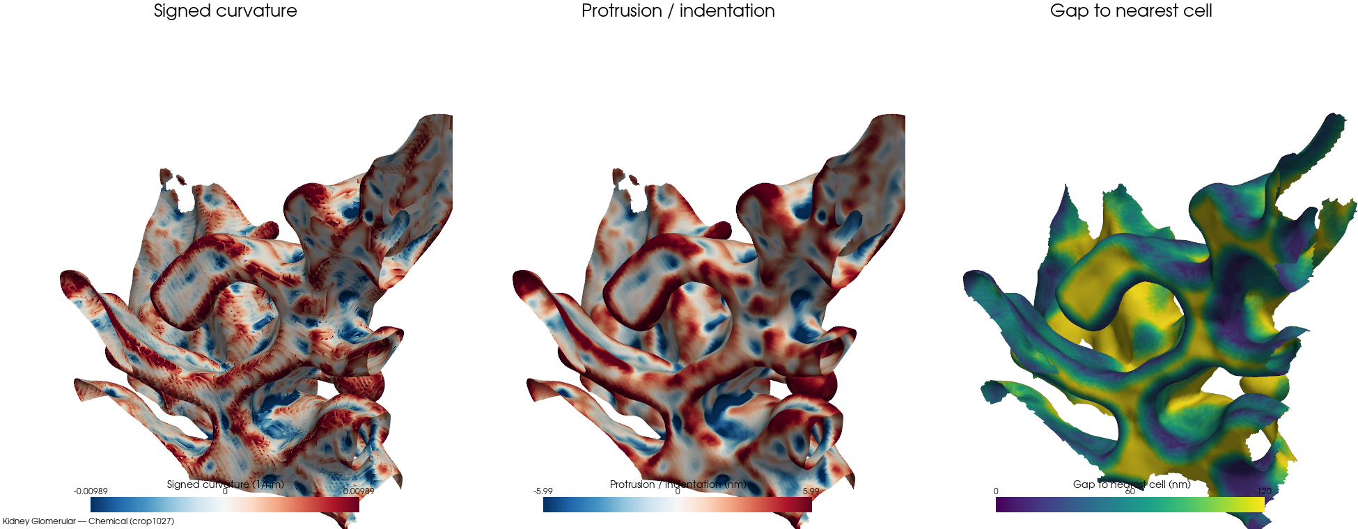
Chemicalcrop1027bd-clip 22%cell 1 · ECS frac 0.4451 · patch 129,021/172,284 faces (74% ECS-facing) · 27 cells · 16 nm · Glomerular interstitium
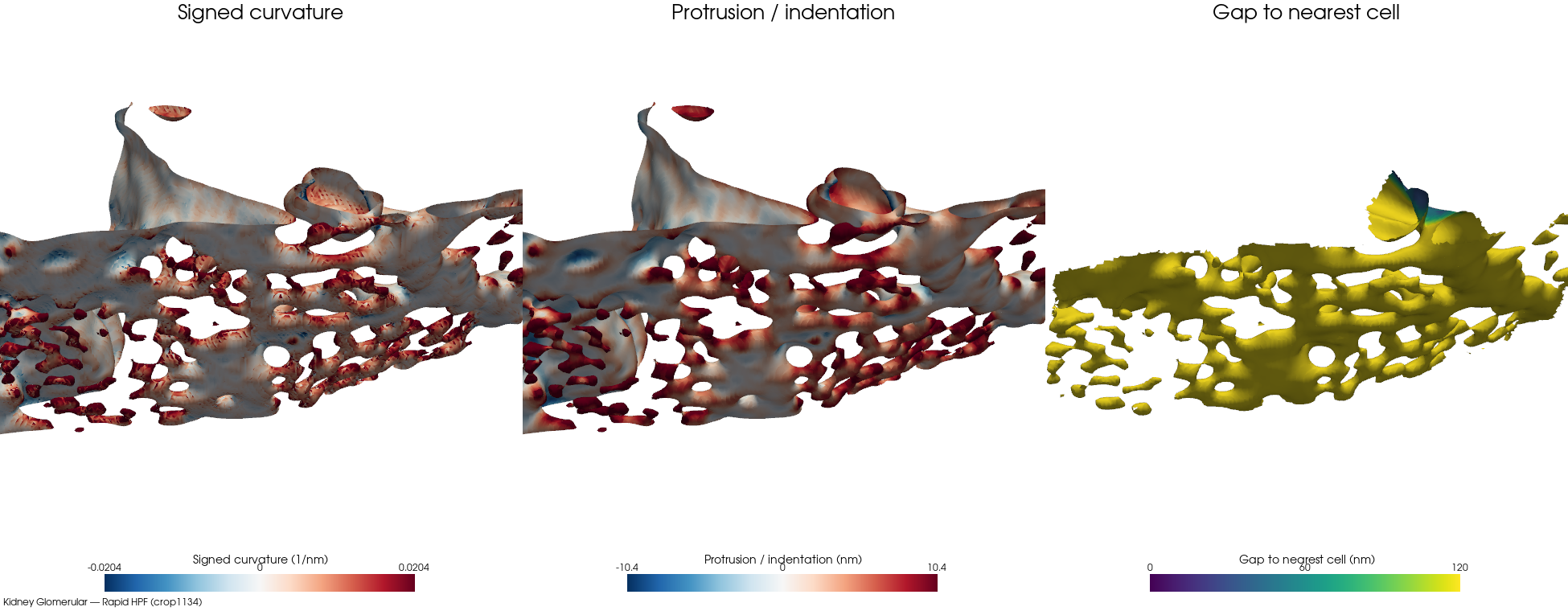
Rapid HPFcrop1134bd-clip 48%cell 1 · ECS frac 0.6465 · patch 129,094/174,494 faces (74% ECS-facing) · 31 cells · 16 nm · Glomerular interstitium
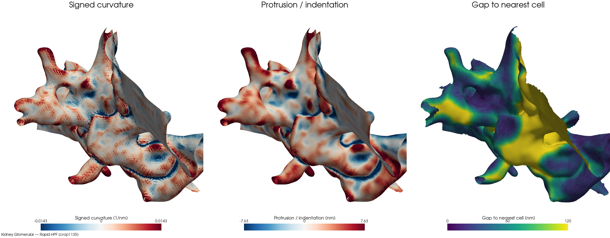
Rapid HPFcrop1135bd-clip 15%cell 1 · ECS frac 0.3478 · patch 98,984/146,308 faces (67% ECS-facing) · 24 cells · 16 nm · Glomerular interstitium
PCT base
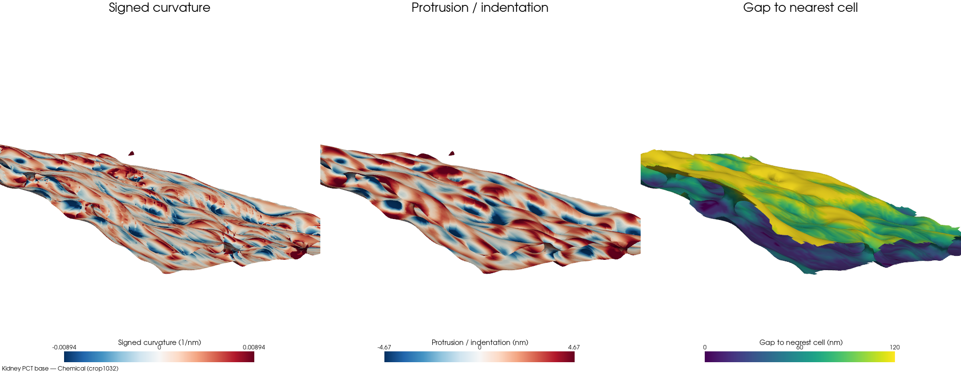
Chemicalcrop1032bd-clip 15%cell 10 · ECS frac 0.3021 · patch 55,646/67,136 faces (82% ECS-facing) · 25 cells · 16 nm · PCT base
PCT brush border
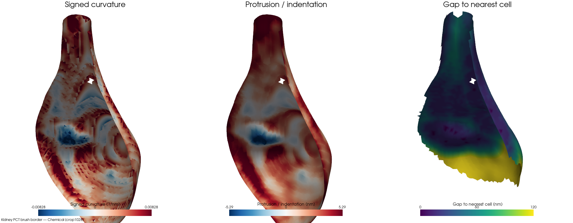
Chemicalcrop1028bd-clip 28%cell 79 · ECS frac 0.5964 · patch 10,394/16,448 faces (63% ECS-facing) · 104 cells · 16 nm · PCT brush border
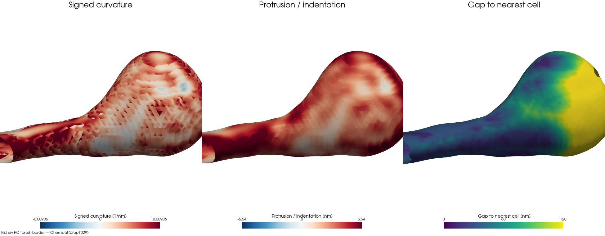
Chemicalcrop1029bd-clip 1%cell 34 · ECS frac 0.7455 · patch 10,170/10,656 faces (95% ECS-facing) · 97 cells · 16 nm · PCT brush border
PCT lateral
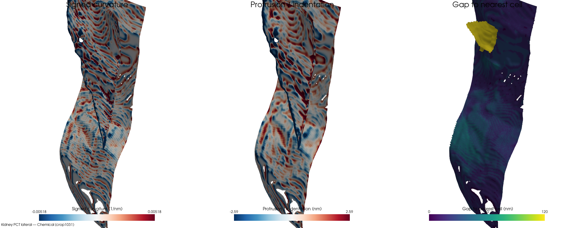
Chemicalcrop1031bd-clip 30%cell 2 · ECS frac 0.0143 · patch 97,903/225,472 faces (43% ECS-facing) · 2 cells · 16 nm · PCT lateral
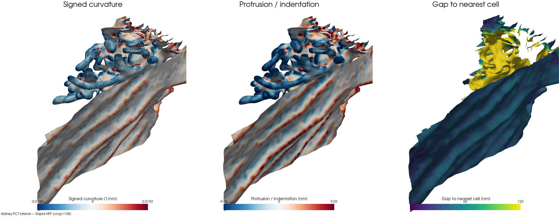
Rapid HPFcrop1138bd-clip 41%cell 2 · ECS frac 0.0898 · patch 306,404/452,202 faces (67% ECS-facing) · 31 cells · 16 nm · PCT lateral
Cortex
(unassigned)
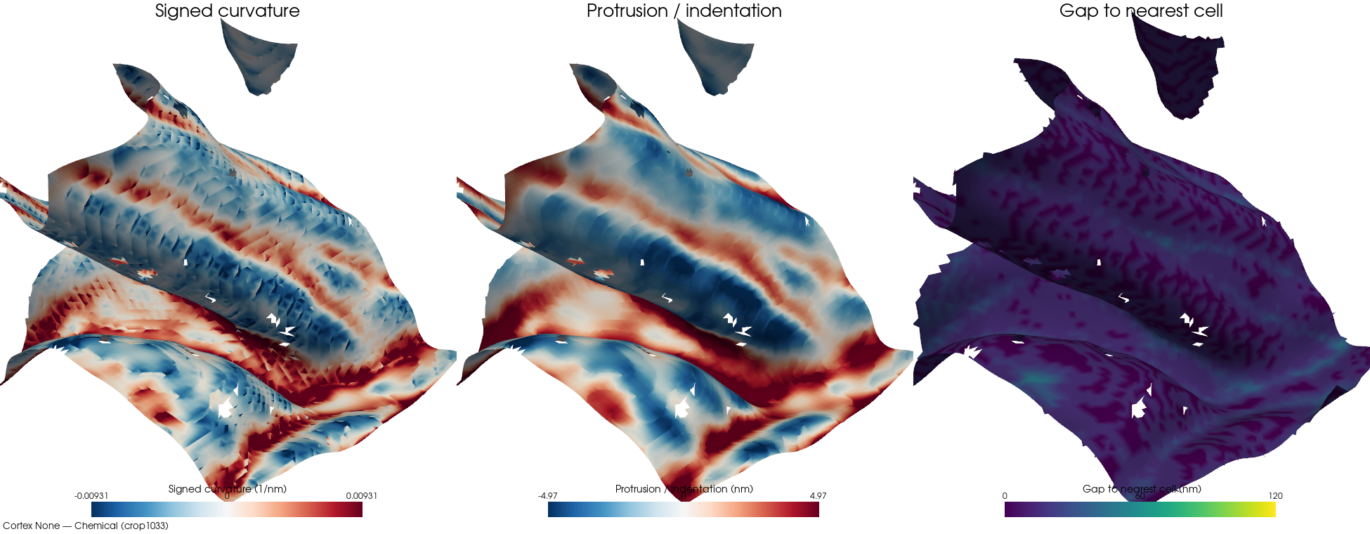
Chemicalcrop1033bd-clip 0%cell 2 · ECS frac 0.0477 · patch 19,700/47,000 faces (41% ECS-facing) · 22 cells · 16 nm · Unmyelinated nerve fibers
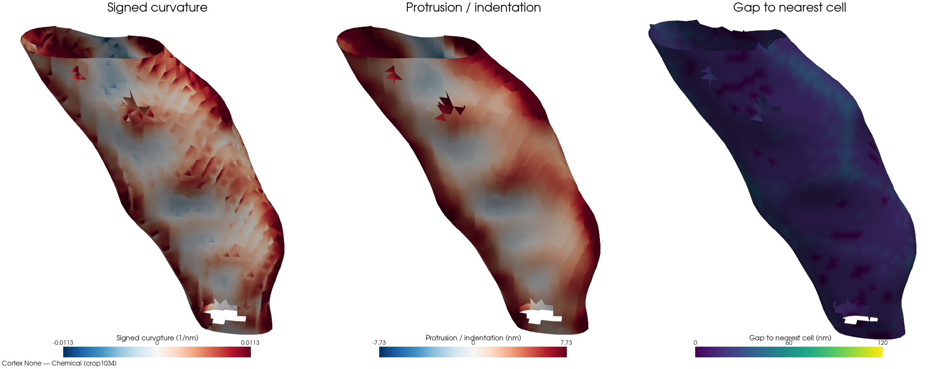
Chemicalcrop1034bd-clip 0%cell 15 · ECS frac 0.0867 · patch 7,060/7,908 faces (89% ECS-facing) · 24 cells · 16 nm · Neuropil (synaptic cleft)
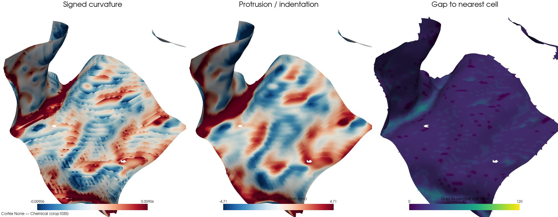
Chemicalcrop1035bd-clip 0%cell 4 · ECS frac 0.0433 · patch 15,431/39,180 faces (39% ECS-facing) · 20 cells · 16 nm · Neuronal cell processes
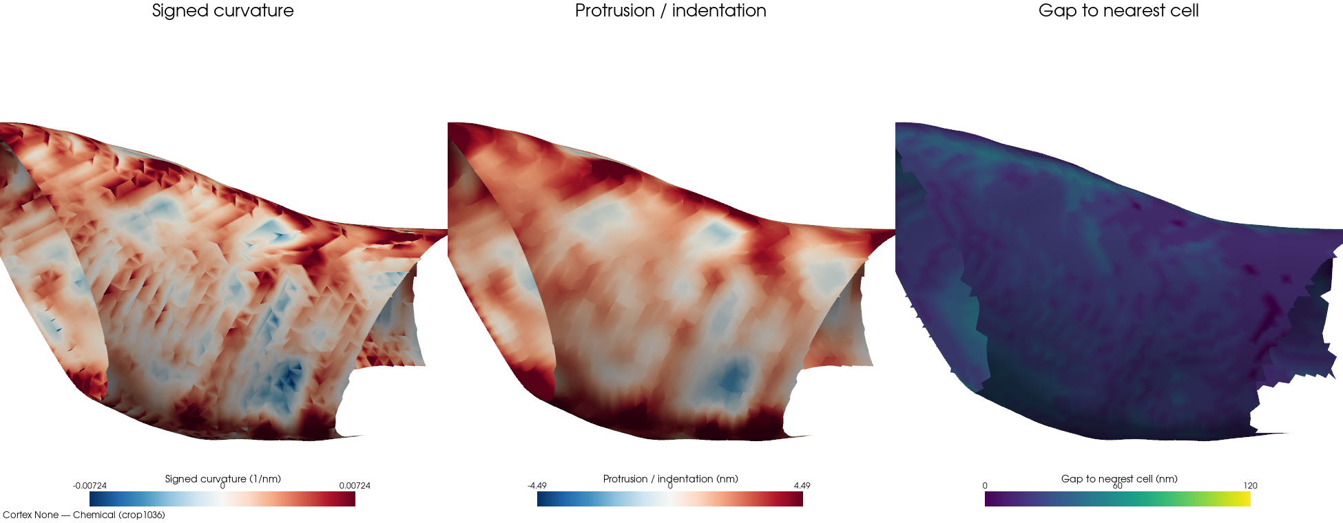
Chemicalcrop1036bd-clip 0%cell 12 · ECS frac 0.1071 · patch 12,206/15,804 faces (77% ECS-facing) · 14 cells · 16 nm · Glial-neuronal (synaptic clefts)
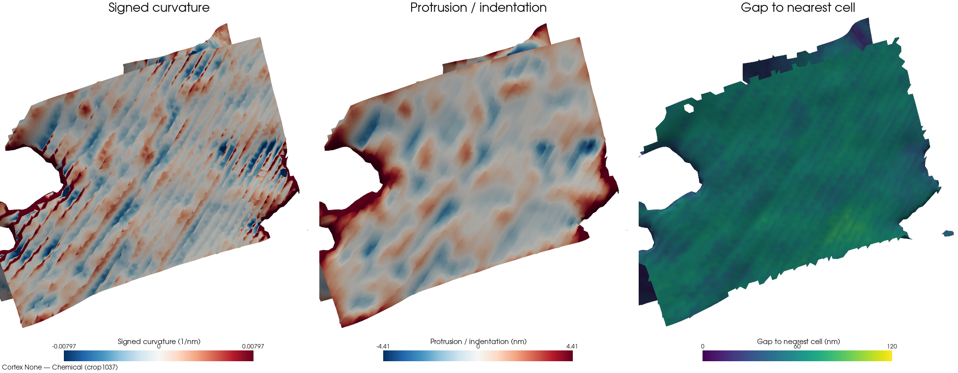
Chemicalcrop1037bd-clip 7%cell 4 · ECS frac 0.3382 · patch 17,515/23,882 faces (73% ECS-facing) · 16 cells · 16 nm · Neurovascular (endothelium/glia/neurons)
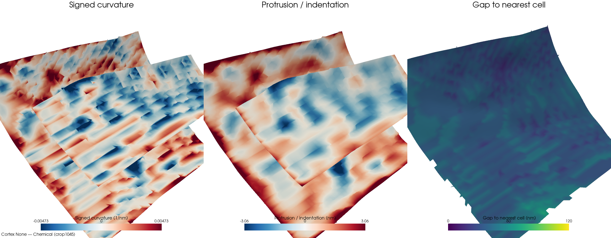
Chemicalcrop1045bd-clip 34%cell 13 · ECS frac 0.1469 · patch 9,177/16,960 faces (54% ECS-facing) · 24 cells · 16 nm · Neurovascular (mixed nerve fibers)
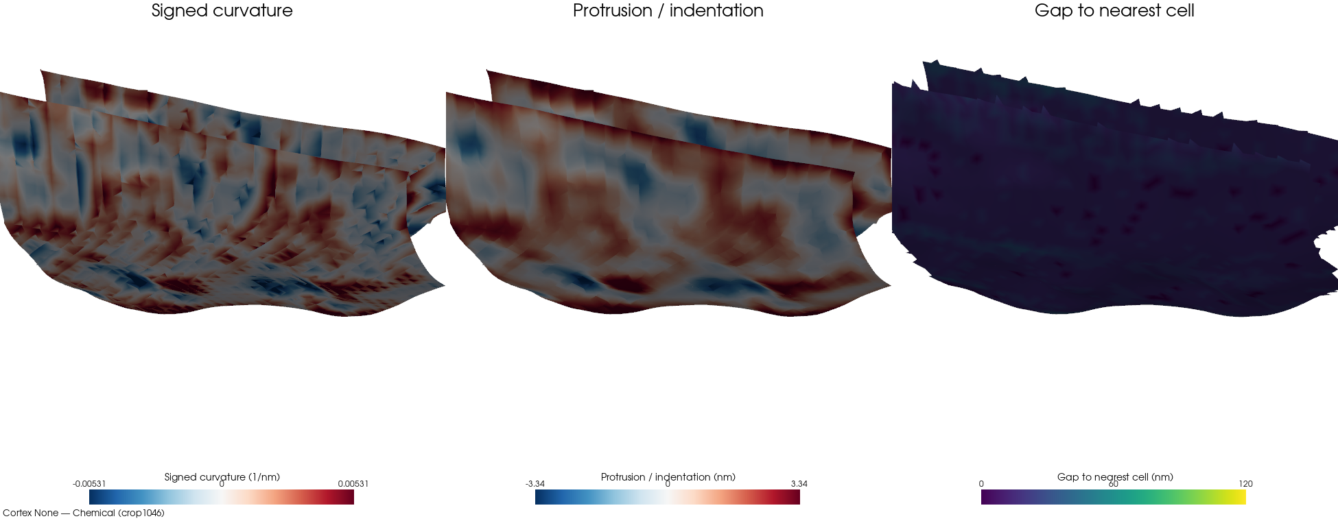
Chemicalcrop1046bd-clip 0%cell 1 · ECS frac 0.1665 · patch 11,343/16,552 faces (68% ECS-facing) · 17 cells · 16 nm · Neurovascular (myelinated/unmyelinated fibers)
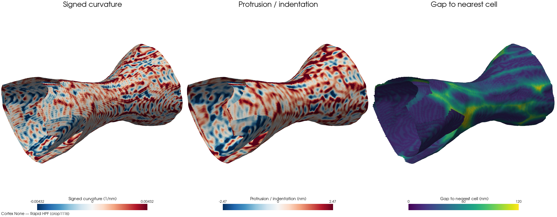
Rapid HPFcrop1116bd-clip 1%cell 62 · ECS frac 0.1995 · patch 193,740/209,900 faces (92% ECS-facing) · 234 cells · 16 nm
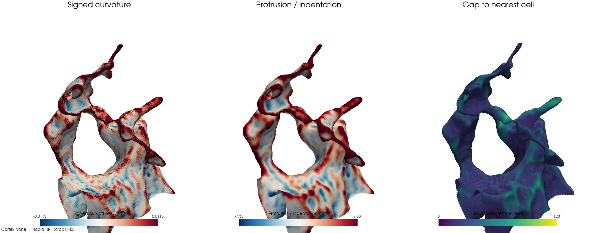
Rapid HPFcrop1140bd-clip 0%cell 31 · ECS frac 0.1471 · patch 82,061/100,400 faces (81% ECS-facing) · 191 cells · 16 nm · Neuropil (unmyelinated fibers)
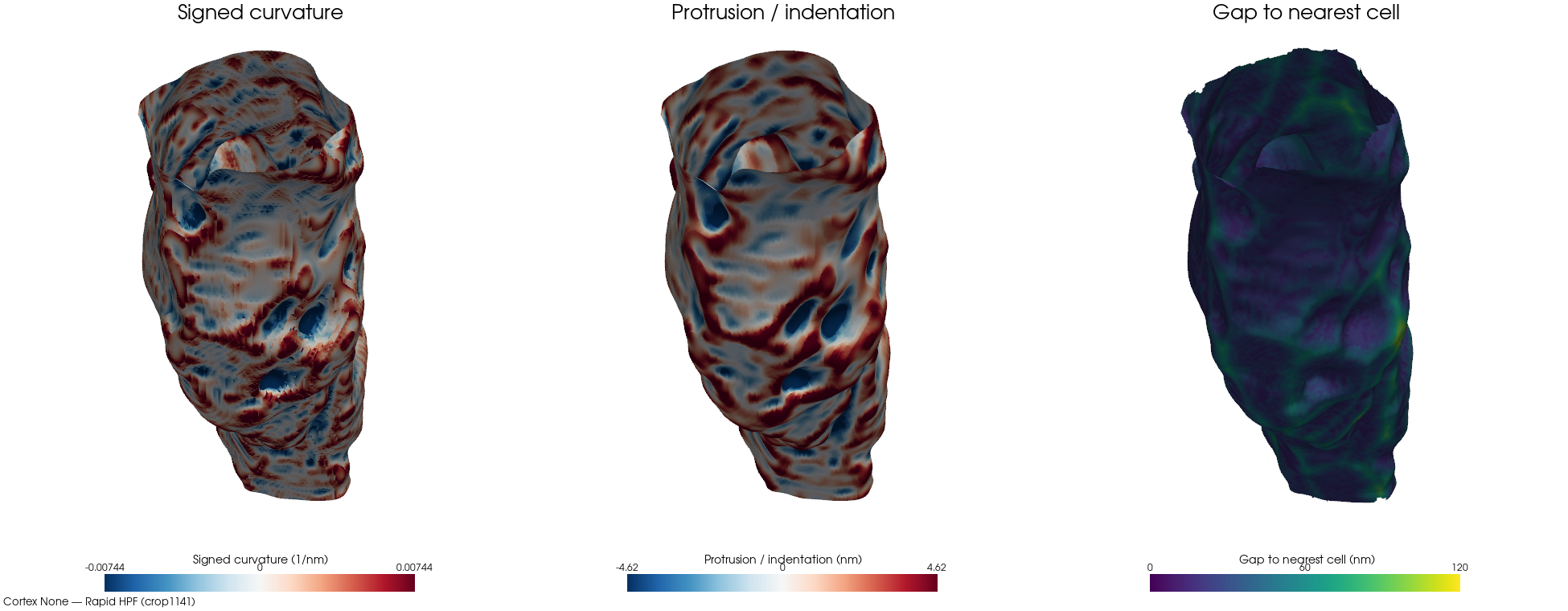
Rapid HPFcrop1141bd-clip 0%cell 13 · ECS frac 0.0959 · patch 106,668/116,828 faces (91% ECS-facing) · 119 cells · 16 nm
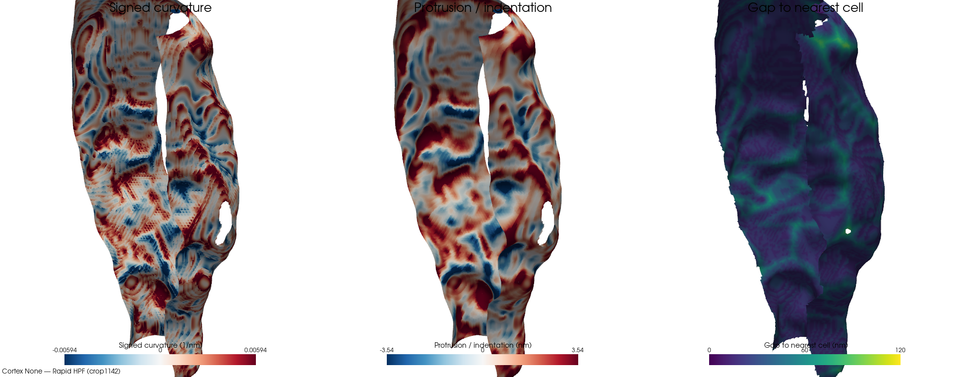
Rapid HPFcrop1142bd-clip 1%cell 51 · ECS frac 0.1248 · patch 61,341/100,968 faces (60% ECS-facing) · 156 cells · 16 nm · Unmyelinated nerve fibers
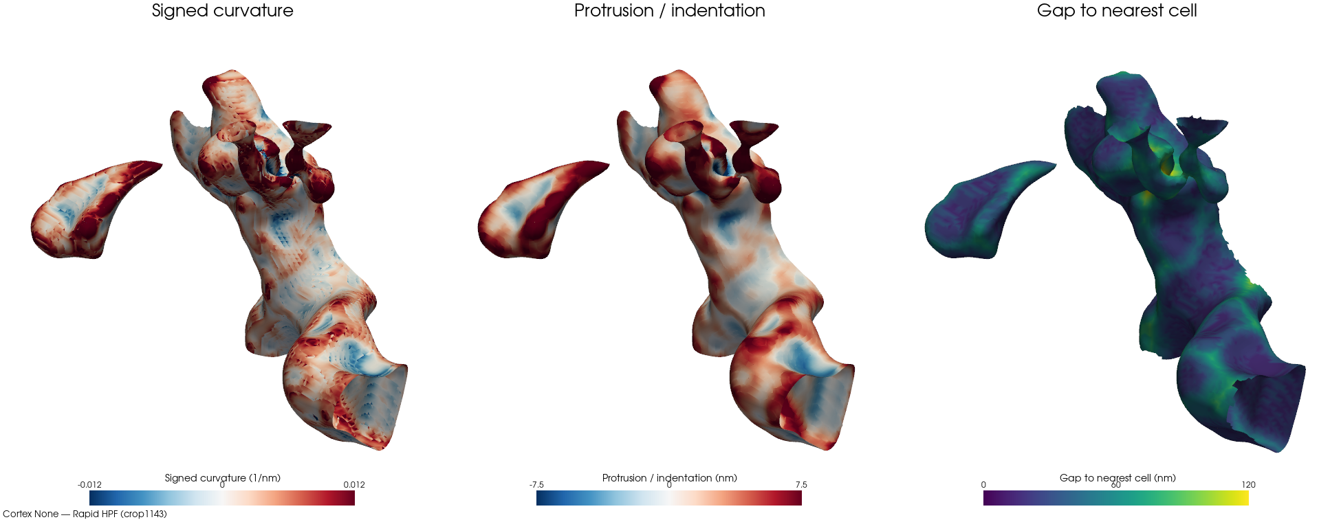
Rapid HPFcrop1143bd-clip 1%cell 13 · ECS frac 0.1815 · patch 47,939/61,080 faces (78% ECS-facing) · 133 cells · 16 nm · Unmyelinated nerve fibers